Workstation
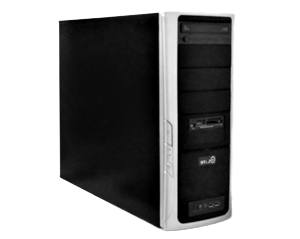
Workstation 1
|
|
CPU: Intel® Core™ 2 QUAD CPU Q9400 @ 2,666 GHz RAM: 3 GB Graphics: NVIDIA GeForce GT 240 – 1024 MB OS: Windows XP Professional Version 2002 HDD space: 368 + 98 GB Software: MetaMorph V.7.7.0; ImageJ/Fiji; FlowLogic Screen: Samsung B2230W – 15.6” - 1360 x 768 @60Hz Booking: Agendo Location: 5.23 Contact: Carolina Feliciano (cfeliciano@itqb.unl.pt) |
Warnings
- Make sure to save all the data to your own storage device or cloud as we routinely wipe out the computer’s data.
- Do not forget to register your utilization in the logbook.
- This workstation is solely for image analysis
Brief Description
The workstation 1 is equipped with MetaMorph software, version 7.7.0, and ImageJ/Fiji. Both software allow the analysis and downstream processing of images acquired with in-house microscopes. This workstation is indicated for several protocols of 2D image analysis and quantification such as phenotype quantification and reporting fluorescence signal expression by using the referred software. Note that this workstation is also available for downstream analysis of flow cytometry data generated by the in-house cell sorter.
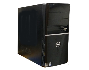
Workstation 2
|
|
CPU: 13th Gen Intel® Core™ I9 @ 2.00 GHz RAM: 64 GB Graphics: NVIDIA T1000 OS: Windows 11 Pro HDD space: 4 TB Software: Zeiss ZEN lite, ImageJ/Fiji, LasX, Imaris Viewer, Cell Profiler, BiofilmQ Screen: Dell E248 WFP - 24” - 1920 x 1200 @60Hz Booking: Agendo Location: 5.23 Contact: Carolina Feliciano (cfeliciano@itqb.unl.pt) |
Warnings
- Make sure to save all the data to your own storage device or cloud as we routinely wipe out the computer’s data
- Do not forget to register your utilization in the logbook
- This workstation is solely for image analysis
Brief Description
The workstation 2 is equipped with ZEISS ZEN lite (supports CZI file format), ImageJ/Fiji, LasX (supports LIF file format), Imaris Viewer, Cell Profiler and BiofilmQ. Softwares allow the analysis and downstream processing of images acquired with in-house microscopes. This workstation is indicated for visualization and analysis of microscope images in 2D and 3D .


