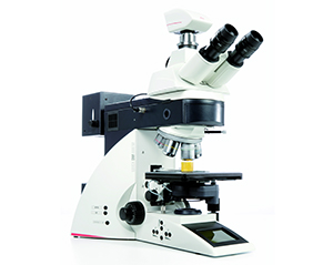
Leica DM 6000B
Widefield Upright Microscope
Transmitted LightPhase Contrast; Bright Field; Dark Field
Fluorescence Filters ForDAPI; CFP; GFP; FITC; YFP; CFP-YFP; CY3; TX2; mCherry
Advanced TechniquesFluorescence recovery after photobleaching (FRAP)Förster resonance energy transfer (FRET)
Training is required before useAn equipment guide is available in our Guides sectionContact BIC for trainingSystem BookingLocation: 5.25A |
|
Warnings
- Before you get started for the first time in this microscope consult the responsible people for proper training
- ONLY turn ON the lamp if it has been off for more than one hour
- Do not turn OFF the lamp if someone else will be using the microscope in the next few hours. To avoid unnecessary working hours or usage constraints, please contact the next user after using the microscope
- Turn ON THE LAMP FIRST and then the remaining devices
- THE LAMP must be the LAST TO TURN OFF
- When closing the MetaMorph software, WAIT for the warning message and let the camera cooling down. In other words, close MetaMorph and DO NOT close any other window that will pop-up until the software closes by itself
- Never LOOK DIRECTLY into the beam paths as they are capable of permanently damaging the human eye
- DO NOT FORGET to clean the oil immersion objectives. Use ONLY the specific tissues (Kimtech®) for this purpose
Brief Description
A fully automated upright microscope equipped with motorized z-focus, coded 7x nosepiece, transmitted light axis and incident light axis. The Leica DM6000 B provides the main transmitted light contrast methods and a user-friendly interface for fluorescence microscopy. Furthermore, the microscope is equipped with a laser unit emitting UV radiation which allows performing the FRAP and FRET techniques.
Suggestion for “Materials and Methods”
Images were acquired on a Leica DM 6000B upright microscope equipped with an Andor iXon 885 EMCCD camera and controlled with the MetaMorph V5.8 software, using the 100x 1.4 NA oil immersion objective plus a 1.6x optvar, the fluorescence filter sets GFP + TX2 and Contrast Phase optics.
Suggestion for “Acknowledgements”
This work was partially supported by PPBI - Portuguese Platform of BioImaging (PPBI-POCI-01-0145-FEDER-022122) co-funded by national funds from OE - "Orçamento de Estado" and by european funds from FEDER - "Fundo Europeu de Desenvolvimento Regional".
Filter Sets
|
Filter Cube |
Excitation Filter |
Dichromatic Mirror |
Emission Filter |
Excitation Colour |
Emission Colour |
|
A4; DAPI1;2 |
BP 360/40 |
400 |
BP 470/40 |
UV |
Blue |
|
CFP |
BP 436/20 |
455 |
BP 480/40 |
Violet/Blue |
Blue/Cyan |
|
L5; GFP1; FITC1;2 |
BP 480/40 |
505 |
BP 527/30 |
Blue/Cyan |
Green |
|
YFP |
BP 500/20 |
515 |
BP 535/30 |
Green |
Green/Yellow |
|
CFP-YFP3 |
BP 436/12 - 500/20 |
445 - 515 |
BP 467/37 - 545/45 |
Violet/Green - Green |
Blue/Cyan – Green/Yellow |
|
CY3; TRITC1 |
BP 545/30 |
565 |
BP 610/75 |
Green/Yellow |
Orange/Red |
|
TX2; FM 4-641; mCherry1 |
BP 560/40 |
595 |
BP 645/75 |
Green/Yellow |
Red |
1 Almost equivalent to this filter cube
2The filter cube may appear entitled to such name in spite of the less known corresponding filter set
3Filter set for the FRET technique
BP stands for a bandpass filter
Objectives
|
Magnification1;2 |
10x |
40x |
63x |
100x |
|
Leica System |
HC |
HCX |
HCX |
HCX |
|
Class |
PL APO (Apochromats) |
PL Fluotar (Semi -Apochromats) |
PL APO (Apochromats) |
PL APO (Apochromats) |
|
Aperture (A) |
0.4 |
0.75 |
1.4 |
1.4 |
|
Immersion |
Air |
Air |
Oil |
Oil |
|
Free Working Distance (mm) |
2.2 |
0.4 |
0.1 |
0.09 |
|
Cover Glass (mm) |
0.17 |
0.17 |
0.17 |
0.17 |
|
Contrasting Methods |
Phase Contrast (PH1) |
Phase Contrast (PH2) |
Phase Contrast (PH3) |
Phase Contrast (PH3) |
|
Pixel size w/ 1.6x optovar (µm)3 |
0.5 | 0.125 | 0.08 | 0.05 |
|
Pixel size w/ 1.25x optovar (µm)3 |
0.64 | 0.16 | 0.1 | 0.064 |
|
Pixel size without optovar (µm)3 |
0.8 | 0.2 | 0.126 | 0.08 |
1Notice that the microscope is equipped with two Eyepieces: HC Plan S 10x/25 Br. M
2Notice that the microscope is equipped with two optvar options: 1.25x; 1.6x
3Pixel sizes were calculated for an Andor IXon EM 885 EMCCD camera
Camera
- Brand: Andor Technology®
- Model: iXon EM+ 885 EMCCD
- Reference: DU 885 K-CSO-#VP
- EMCCD Technology
- 1004 x 1002 active pixels
- 4 Frame Rate @ full resolution (frames per sec)
- 8 x 8-pixel size (WxH; µm)

