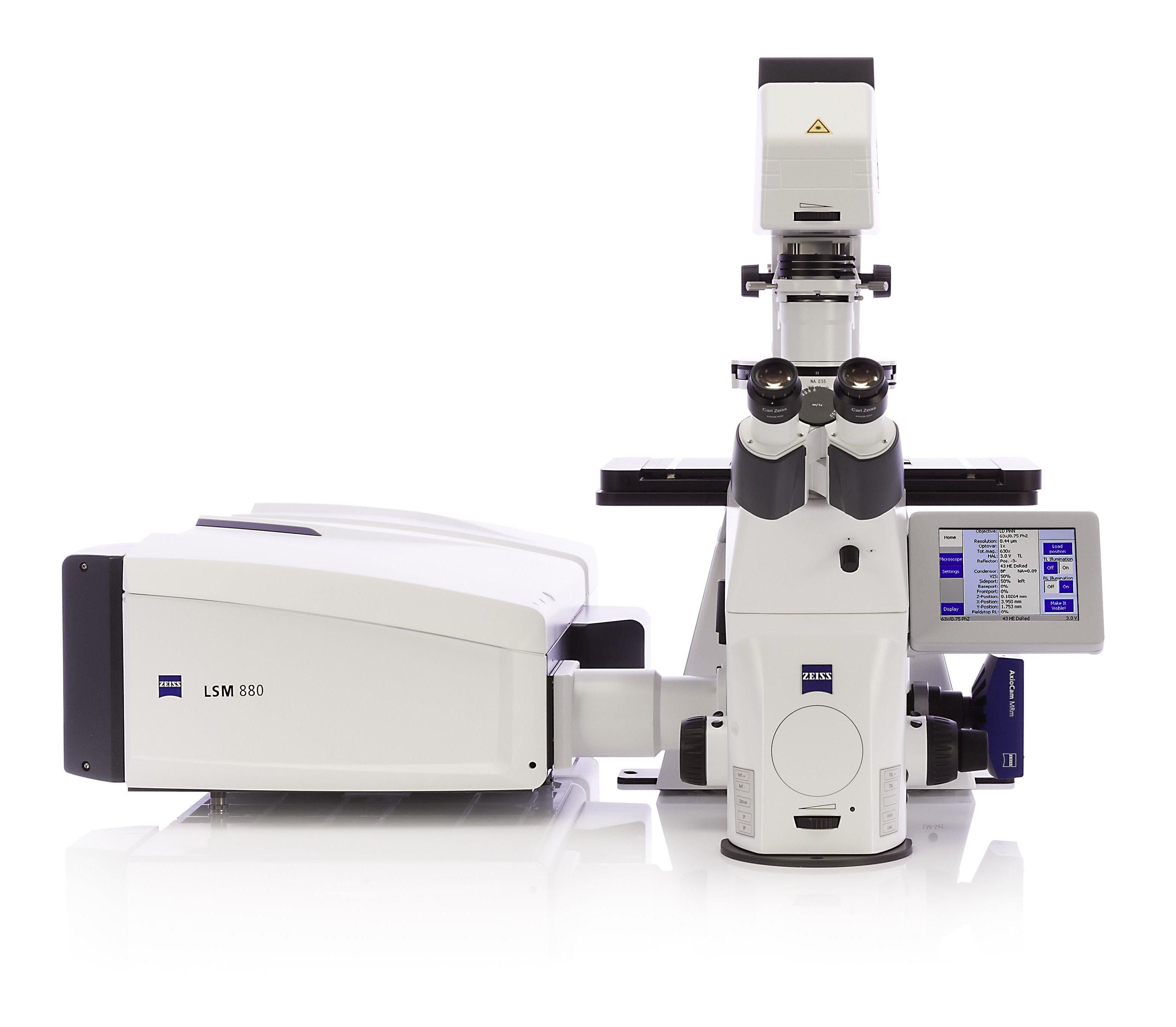
Zeiss LSM 880 w/ Fast Airyscan
Inverted Laser Scanning Confocal Microscope
Laser lines405 nm; 458 nm; 488 nm; 514 nm; 561 nm; 633 nm
Detectors2 PMTs; 1 GaAsP PMT; 1 T-PMT; 1 Airyscan Detector with Fast Module
Transmitted LightBright Field; Differential Interference Contrast (DIC)
Available TechniquesZ-stacks; Multiple Positions; Time series; Tile scanSuper Resolution Imaging with the Airyscan detector
Training is required before useAn equipment guide is available in our Guides sectionContact BIC for trainingSystem BookingLocation: 5.01 |
|
Warnings
-
The equipment cannot be used without official training
-
Before your first use contact the responsible people to schedule training
-
Never LOOK DIRECTLY into the beam paths as they are capable of permanently damaging the human eye
-
Save all data into your own storage device as we routinely clean the computer’s data
-
Register your utilization in the logbook
-
Airyscan raw data reconstruction must be done in the microscope computer (book slots accordingly). For all downstream image analysis image analysis a free software license is available for ZEN lite at ZEISS website. Alternatively, you can use FIJI.
-
Contact the responsible people in case of any doubt or any issue with the equipment
Booking Rules
Booking is done online at the ITQB intranet. Please mind the following rules when booking the microscope:
- Priority booking of afternoon slots is given to users with live cell experiments that require growth in the same day. If you have fixed or time insensitive samples, please try to book other times;
- Maximum 2 slots per user per week (one slot = one morning or one afternoon);
- These rules are applicable until the previous Friday. If you need more than two slots in a week and they are still available on the previous Friday, at 4pm, feel free to book them.
Know Your Fluorophores
Use resources such as the FPBase or the Thermofisher SpectraViewers to explore your fluorophores. Learn their excitation and emission wavelengths, and what are their optimal laser lines and filters. This is necessary to use the confocal to its full potential.
Brief Description
Zeiss LSM 880 is an inverted confocal laser scanning microscope with Airyscan, a new detector concept featuring a 32 channel GaAsP-PMT area detector. The Airyscan detector thus brings forward simultaneous improvements in spatial resolution and signal-to-noise ratio in comparison with conventional confocal microscopy. This allows the LSM 880 to resolve small structures up to 120 nm (XY) and 350 nm (Z) even in thick samples.
The introduction of a Fast module for the Airyscan offers a satisfactory compromise between perfect optical sections and live cell imaging experiments. The Fast Airyscan mode reduces photo-bleaching and allows acquisition speeds of up to 19 frames per second at 512 x 512-pixel image resolution.
The LSM 880 with Airyscan highlights the needs of your research for quantitative data in live cell experiments at superior resolutions.
Suggestion for “Materials and Methods”
Confocal Z-series stacks (step size 200 nm) were acquired on a Zeiss LSM 880 point scanning confocal microscope using the Airyscan detector, a 63x Plan-Apochromat 1.4NA DIC oil immersion objective (Zeiss) and the 405 nm, 488 nm and 561 nm laser lines. The Zeiss Zen 2.3 (black edition) software was used to control the microscope, adjust spectral detection for the emission of 4′,6-diamidino-2-phenylindole (DAPI), Green Fluorescent Protein (GFP) and tetramethylrhodamine (TMR) SNAP-tag fluorochromes and for processing of the Airyscan raw images.
Confocal Z-series stacks and tile regions with multiple positions were acquired on a Zeiss LSM 880 point scanning confocal microscope using photomultiplier tube detectors (PMTs) and a gallium arsenide phosphide (GaAsP) detector, a 63x Plan-Apochromat 1.4NA DIC oil immersion objective (Zeiss) and the 405 nm, 488 nm and 561 nm laser lines. The Zeiss Zen 2.3 (black edition) software was used to control the microscope and adjust spectral detection for the emission of 4′,6-diamidino-2-phenylindole (DAPI), Green Fluorescent Protein (GFP) and tetramethylrhodamine (TMR) SNAP-tag fluorochromes.
Images were acquired on a Zeiss LSM 880 point scanning confocal microscope controlled with the Zeiss Zen 2.3 (black edition) software, using a 63x Plan-Apochromat 1.4NA DIC oil immersion objective (Zeiss), the fluorescence filter sets GFP + TX2 and differential interference contrast (DIC) optics.
Information to be added to the Acknowledgements section
This work was partially supported by PPBI - Portuguese Platform of BioImaging (PPBI-POCI-01-0145-FEDER-022122) co-funded by national funds from OE - "Orçamento de Estado" and by european funds from FEDER - "Fundo Europeu de Desenvolvimento Regional".
Filter Sets for Incident Light
|
Filter Cube |
Excitation Filter |
Dichromatic Mirror |
Emission Filter |
Emission Colour |
|
DAPI |
G 365 |
395 |
BP 445/50 |
Blue |
|
GFP |
BP 470/40 |
495 |
BP 525/50 |
Green |
|
Cy3.5 |
BP 565/30 |
585 |
BP 620/60 |
Orange/Red |
BP stands for a bandpass filter
Objectives
|
Magnification1 |
10x |
20x |
40x2 |
63x |
|
ZEISS System |
EC Plan-Neofluar |
Plan-Apochromat |
Plan-Apochromat |
Plan-Apochromat |
|
Class |
Colour and Chromatic Correction |
Colour and Chromatic Correction |
Colour and Chromatic Correction |
Colour and Chromatic Correction |
|
Numerical Aperture (NA) |
0.3 |
0.8 |
1.2 |
1.4 |
|
Immersion |
Air |
Air |
Water3, Oil and Glycerine |
Oil |
|
Free Working Distance (mm) |
5.2 |
0.55 |
0.41 |
0.19 |
|
Cover Glass (mm) |
0.17 |
0.17 |
0.17 |
0.17 |
|
Contrasting Methods |
None |
Differential Interference Contrast (DICII) |
Differential Interference Contrast (DIC) |
Differential Interference Contrast (DICIII) |
1Notice that the microscope is equipped with two Eyepieces: HC Plan S 10x/25 Br. M
2Objective only available by request
3

