Cell handling/Cell characterisation
In the past few years, there has been extensive research in the manipulation and analysis of biological cells at the microscale and a number of microelectromechanical systems (MEMS) for selective manipulation or identification of cells were described. The goal has been the development of automated single-cell manipulation and analysis systems to be used in research fields such as immunology, developmental biology, tumour biology and parasitology.
Dielectrophoresis (DEP)
DEP is a well known method for manipulation of dielectric particles such as DNA, proteins and cells. It is defined as the translational motion impaired on uncharged cells or particles, as a result of the polarisation induced by non-uniform electric fields. The dielectrophoretic force is dependent on the dielectric properties and size of the cell, frequency of the applied electric field and conductivity and permittivity of the medium where the cells are suspended. The selection of these parameters can induce cell movement in specific directions according to their dielectrical properties, thus enabling cell manipulation or separation.
Experiments have been made towards the separation of normal and Babesia bovis infected erythrocytes. The electrical properties of the infected cells are altered due to the presence of the parasite, allowing the separation of normal erythrocytes from infected ones by the application of a suitable electric field.
Figure 1 shows the experimental microfabricated chip used in cell separation and the complete setup used in the experiments, including the flow control apparatus and electrical connections. This chip has built-in microchannels through which the cell solution is conducted. Etched metal electrodes are positioned along the main channel, where the electric potentials are applied to induce the DEP force.
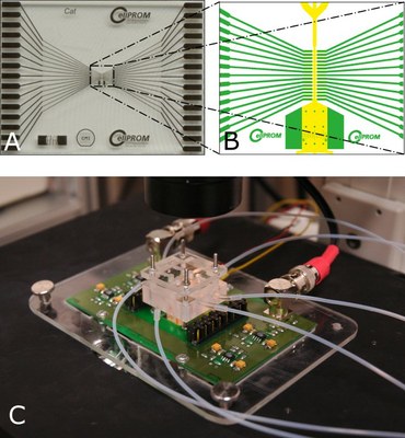
Figure 1 – Microfabricated flow cytometer setup. (A) Chip photograph. (B) Illustration of the electrode pairs along the microchannel. (C) Complete setup, including electrical and pneumatic connections (in collaboration with EPFL/Lausanne-Switzerland - Project CellPROM).
Impedance Spectroscopy (IS)
IS, also known as dielectric spectroscopy, is a noninvasive technique that can be used for characterization of living cells. In addition of quantifying cell concentration, impedance measurements over a wide range of frequencies provide information about cell size, membrane capacitance and cytoplasm properties as a function of frequency. These data can be used to distinguish cell populations without the need for cell markers because cells can be described in terms of their electrical characteristics.
An AC electric potential at a defined set of frequencies is applied to two pairs of electrodes inside a microchannel (Figure 2). This potential applied over each pair of electrodes results in an electric field and every passing cell crossing this electric field modifies temporarily the impedance as well as the current allowing the detection of membrane and cytoplasm properties. The electronic circuit converts the currents in potentials comparing them in order to perform a differential measurement of the impedance between the two electrode pairs to avoid signal drift.
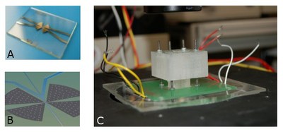
Figure 2 – (A, B) Microfluidic platform with two pair of platinum electrodes and SU-8 channels; (C) Side view of the experimental setup used for IS measurements with a microfluidic platform. (in collaboration with EPFL/Lausanne-Switzerland - Project CellPROM)


